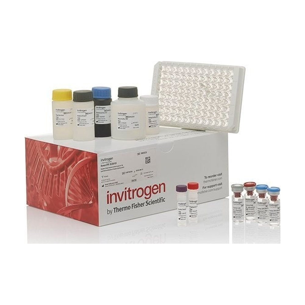
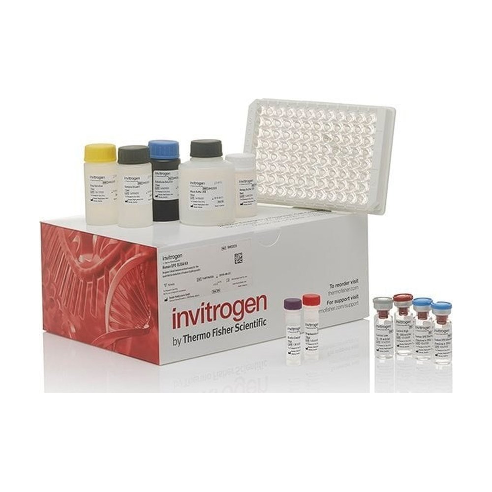
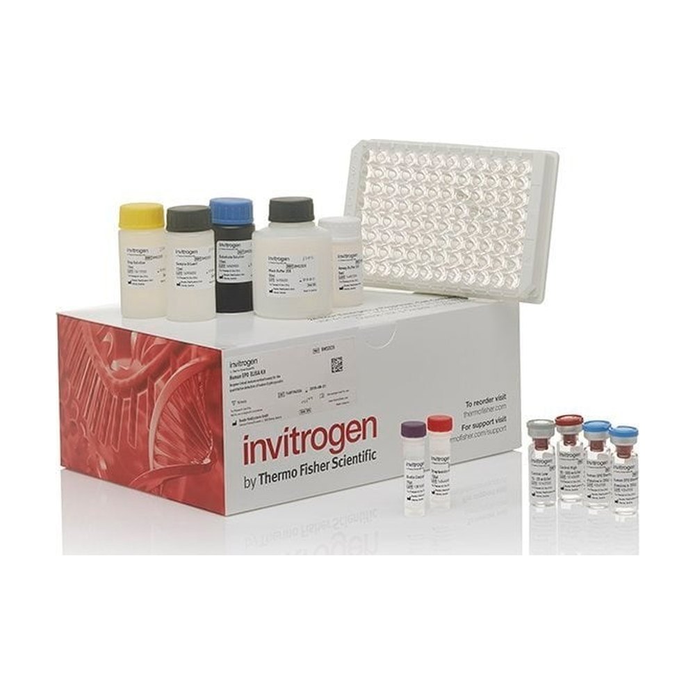
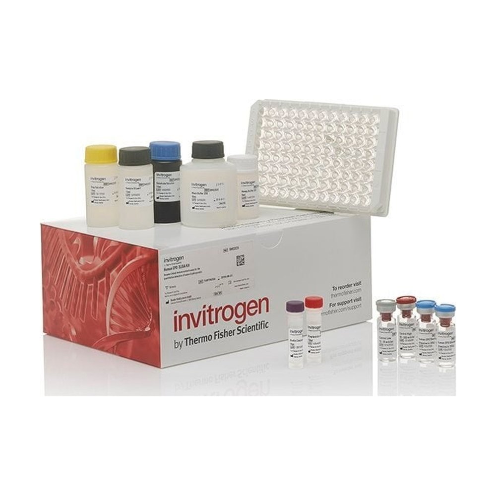
The DNA Damage ELISA quantitates PGFM in serum, plasma, saliva, urine, digested DNA, fecal extracts, or cell culture medium. The assay will exclusively recognize both natural and recombinant DNA Damage. Principle of the method The PGFM solid-phase…
Dose range
62.6-8000 pg/ml
Sensitivity of the dosage
50.9 pg/ml
Description
Competitive ELISA kit for damage to the DNA
Dose
Multiplex strength on Luminex platform
Conjugate
HRP
Brand
Invitrogen
The DNA Damage ELISA quantitates PGFM in serum, plasma, saliva, urine, digested DNA, fecal extracts, or cell culture medium. The assay will exclusively recognize both natural and recombinant DNA Damage. Principle of the method The PGFM solid-phase…
The DNA Damage ELISA quantitates PGFM in serum, plasma, saliva, urine, digested DNA, fecal extracts, or cell culture medium. The assay will exclusively recognize both natural and recombinant DNA Damage. Principle of the method The PGFM solid-phase sandwich ELISA (enzyme-linked immunosorbent assay) is designed to measure the amount of the target bound between a matched antibody pair. A target-specific antibody has been pre-coated in the wells of the supplied microplate. Samples, standards, or controls are then added into these wells and bind to the immobilized (capture) antibody. The sandwich is formed by the addition of the second (detector) antibody, binding to the target on a different epitope from the capture antibody. A conjugated enzyme has been incorporated into the assay. After incubation and washing steps to rid the microplate of unbound substances, a substrate solution is added that reacts with the enzyme-antibody-target complex to produce measurable signal. The intensity of this signal is inversely proportional to the concentration of target present in the original specimen. Rigorous validation Each manufactured lot of this ELISA kit is quality tested for criteria such as sensitivity, specificity, precision, and lot-to-lot consistency. See manual for more information on validation.
Free radicals and other reactive species are constantly generated in vivo and cause oxidative damage to biomolecules, a process that can only be verified by the existence of several antioxidant and repair systems, as well as by the replacement of damaged nucleic acids, proteins and lipids. Intracellular free radical species (ROS) are produced following normal metabolism while extracellular forms are generated by ultraviolet or ionizing radiation. Cell function can be interrupted or stopped if damage caused by the DNA corrupts the integrity of the essential information contained in the genome. When the individual bases are damaged, non-specific repair enzymes of the DNA excise d-DNA lesions to release the deoxynucleotides while the base-specific repair glycosylases excise the corresponding base. Deoxynucleotides are enzymatically hydrolyzed to stable deoxynucleosides. These repair products are transported in the blood and excreted in the urine. The damage to the RNA is reflected in nucleoside adducts. Among many types of oxidative DNA damage, the formation of 8-hydroxy-2-deoxyguanosine (8-OHdG) is a ubiquitous marker of oxidative stress. 8-OHdG is formed physiologically and then improved by chemical carcinogens. When repairing in vivo exonuclease damage to DNA, the 8-OHdG created was then excreted without further metabolism in urine. 8-hydroxy-2-deoxyguanosine (8-OHdG) is identical in all species.
Sensitivity
Sensitivity of the dosage: 50.9 pg/ml.
Measurement range
Dose range: 62.6-8000 pg/ml.
Duration of dosing
Total duration of dosing: 2 h 30 min.
Reproducibility
Coefficient of variation intra-dose 9%, inter-dose 9.9%.
ELISA, protein assays, activity tests, protein biology, reproductive biology, inflammation, oxidative stress, metabolism, cell signaling, epigenetics, haematology, damage to the DNA, cancer biology, immunology, neurosciences, neurobiology, hormone detection s