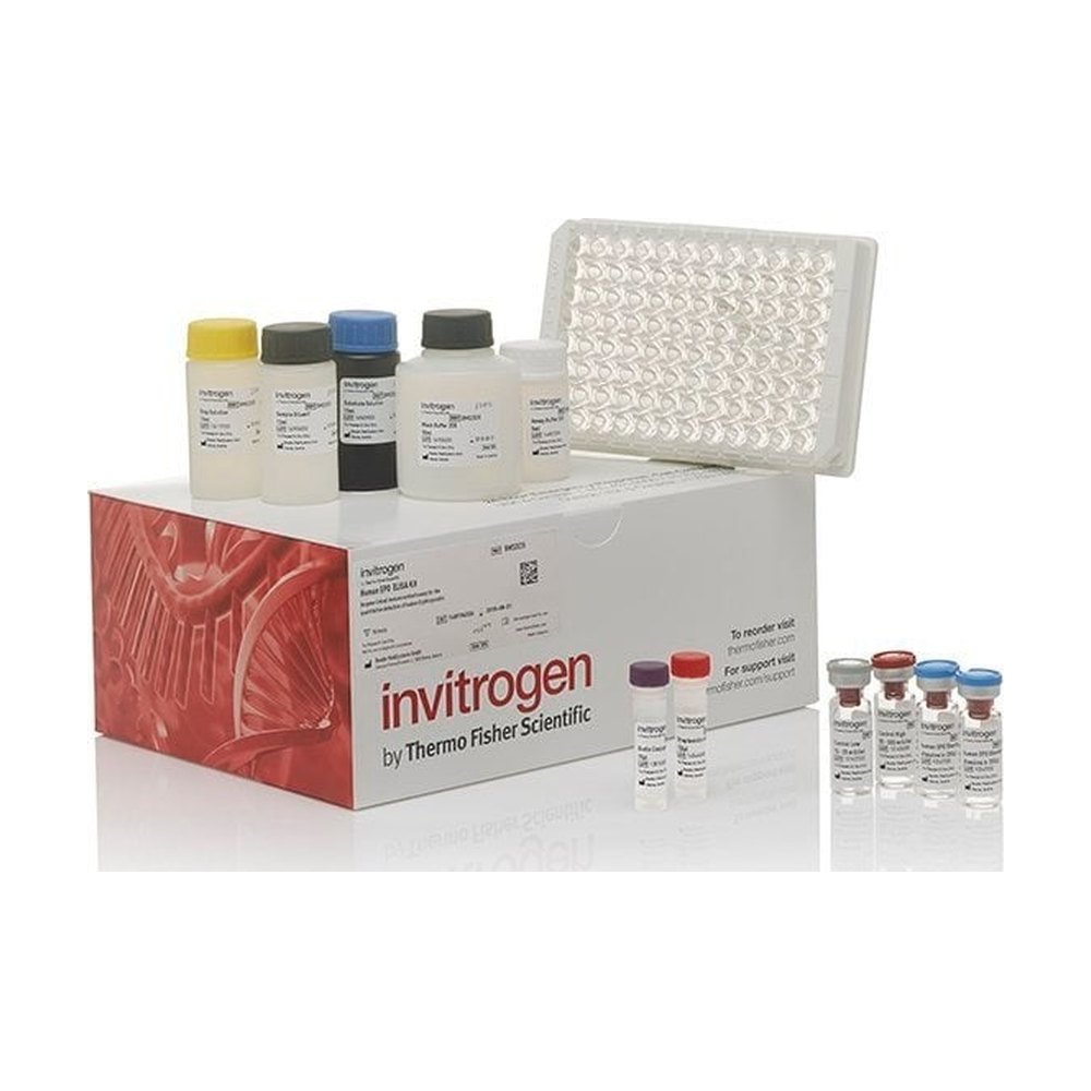
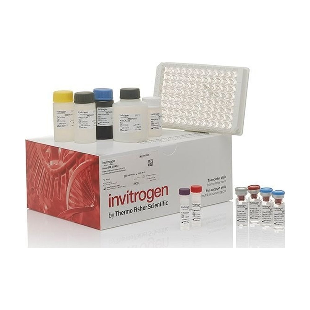
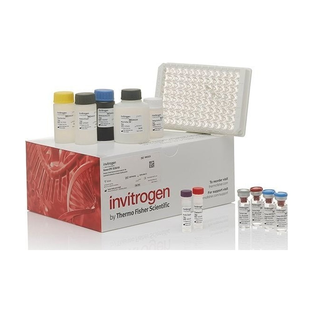
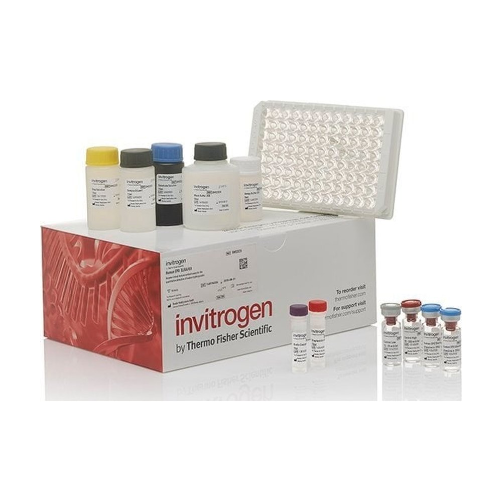
Kit ELISA sandwich PAI-1 (SERPINE1) (singing): quantitative determination of Rhesus Mk PAI-1 in serum, plasma or cell culture medium. The test recognizes only the natural and recombinant forms of Mk PAI-1.
Accession number
XP_001107647.1
Dose range
24.58-6000 pg/ml
Sensitivity of the dosage
20 pg/ml
Calibration range
20 pg/ml to 6.000 pg/ml
Dose
ELISA solid-phase sandwich
Brand
Invitrogen
Kit ELISA sandwich PAI-1 (SERPINE1) (singing): quantitative determination of Rhesus Mk PAI-1 in serum, plasma or cell culture medium. The test recognizes only the natural and recombinant forms of Mk PAI-1.
Kit ELISA sandwich PAI-1 (SERPINE1) (singing): quantitative determination of Rhesus Mk PAI-1 in serum, plasma or cell culture medium. The test recognizes only the natural and recombinant forms of Mk PAI-1.
Method principle: This ELISA solid phase sandwich test (immuno-enzymatic dose) measures the amount of target bound between a pair of matched antibodies. A specific target antibody is pre-adsorbed into the wells of the supplied microplate. Samples, standards or controls are added to these wells and bind to immobilized antibodies (capture antibodies). The sandwich is formed by adding the second antibody (detection antibody), then a substrate solution reacts with the enzyme-antibodies-target complex to produce a measurable signal. The intensity of this signal is directly proportional to the concentration of the target present in the original sample.
Rigorous validation: Each batch manufactured from this ELISA kit is checked for criteria such as sensitivity, specificity, accuracy and reproducibility from one batch to another (see the package leaflet for more information on validation).
PAI1 (plasminogen 1 activator inhibitor) belongs to the superfamily of serine protease inhibitor and is the main inhibitor of tissue-type and urokinase-type plasminogen activators (tPA and uPA). Platelets are the main source of circulating PAI11, but are synthesized and secreted by many types of tissues and cells, including fibroblasts, smooth muscle cells, endothelial cells, hepatocytes and inflammatory cells. The expression of PAI11 can be regulated at the transcriptional level by many factors, including growth factors, cytokines, hormones, inflammatory factors, glucose or lipid metabolites, vascular tone control factors, chemicals, and other environmental or physical factors. IAP1 is present at increased levels in various disease states, and has been associated with an increase in the occurrence of thrombosis in obesity, thrombophilia and metabolic syndrome. IAP-1 defects are characterized by abnormal bleeding. PAI1 media inhibition of fibrinolysis by inhibiting the activity of plasminogenic activator and may promote neuronal survival. Other PAI1 defects are the cause of a deficiency in plasminogen 1 (PAI-1 deficiency) inhibitor. Alternative isoform epistic transcription variants of PAI1 have also been found.