"/> Raw 264.7 (mouse macrophage, Abelson murine leukemia virus tr… | B-LABO France

Raw 264.7 (mouse macrophage, Abelson murine leukemia virus transformed) Nuclear extract lysate, Denatured; Abnova

Réf. : BL-RE-109629Réf. fabricant : 16099565 50 μg
209,00 € HT

À utiliser avec (application)
Western Blot
Tissue
Macrophage
Description
Nuclear extract cell lysate (denatured)
Tampon de lyse
Buffer A: 10mM HEPES pH 7.9, 1.5mM MgCl2, 10mM KCl, 0.5 mM DTT. Buffer C: 20mM HEPES pH 7.9, 25%(v/v) Glycerol , 0.42M NaCl , 1.5mM MgCl2, 0.2 mM EDTA, 0.5 mM DTT & 0.5 mM PMSF. Buffer D : 20mM HEPES pH 7.9, 20%(v/v) glycerol, 50mM KCl, 0.2 mM EDTA, 0.5 mM DTT & 0.5 mM PMSF.
Méthode de préparation
Nuclear extract was prepared by using a modified protocol of Dignam et al. Cells were Harvested and homogenized in Buffer A, and then centrifugated at 25,000 g for 20 minutes to remove cytoplasm and pellet the nuclei. The pellet was re-suspended in Buffer C, and then the suspensions were centrifuged to collect nuclear extract. The supernatant was dialyzed against Buffer D. The dialysate was then centrifuged, divided into aliquots, and stored at -80°C. The protein concentration was determined by the method of Bradford (Bio-Rad protein assay, microplate standard assay). The lysate was adjusted to 2.5 mg/ml, and then mixed with 5X Sample Buffer to become final 2 mg/ml in 1X Sample Buffer. The lysate was heated at 95°C for 5 min, and cooled rapidly.
Marque
Abnova

Western Blotting

Caractéristiques techniques

MarqueAbnova
Réf. B-LABOBL-RE-109629
Réf. fabricant16099565
À utiliser avec (application)Western Blot
TissueMacrophage
Tampon de lyseBuffer A: 10mM HEPES pH 7.9, 1.5mM MgCl2, 10mM KCl, 0.5 mM DTT. Buffer C: 20mM HEPES pH 7.9, 25%(v/v) Glycerol , 0.42M NaCl , 1.5mM MgCl2, 0.2 mM EDTA, 0.5 mM DTT & 0.5 mM PMSF. Buffer D : 20mM HEPES pH 7.9, 20%(v/v) glycerol, 50mM KCl, 0.2 mM EDTA, 0.5 mM DTT & 0.5 mM PMSF.
Méthode de préparationNuclear extract was prepared by using a modified protocol of Dignam et al. Cells were Harvested and homogenized in Buffer A, and then centrifugated at 25,000 g for 20 minutes to remove cytoplasm and pellet the nuclei. The pellet was re-suspended in Buffer C, and then the suspensions were centrifuged to collect nuclear extract. The supernatant was dialyzed against Buffer D. The dialysate was then centrifuged, divided into aliquots, and stored at -80°C. The protein concentration was determined by the method of Bradford (Bio-Rad protein assay, microplate standard assay). The lysate was adjusted to 2.5 mg/ml, and then mixed with 5X Sample Buffer to become final 2 mg/ml in 1X Sample Buffer. The lysate was heated at 95°C for 5 min, and cooled rapidly.
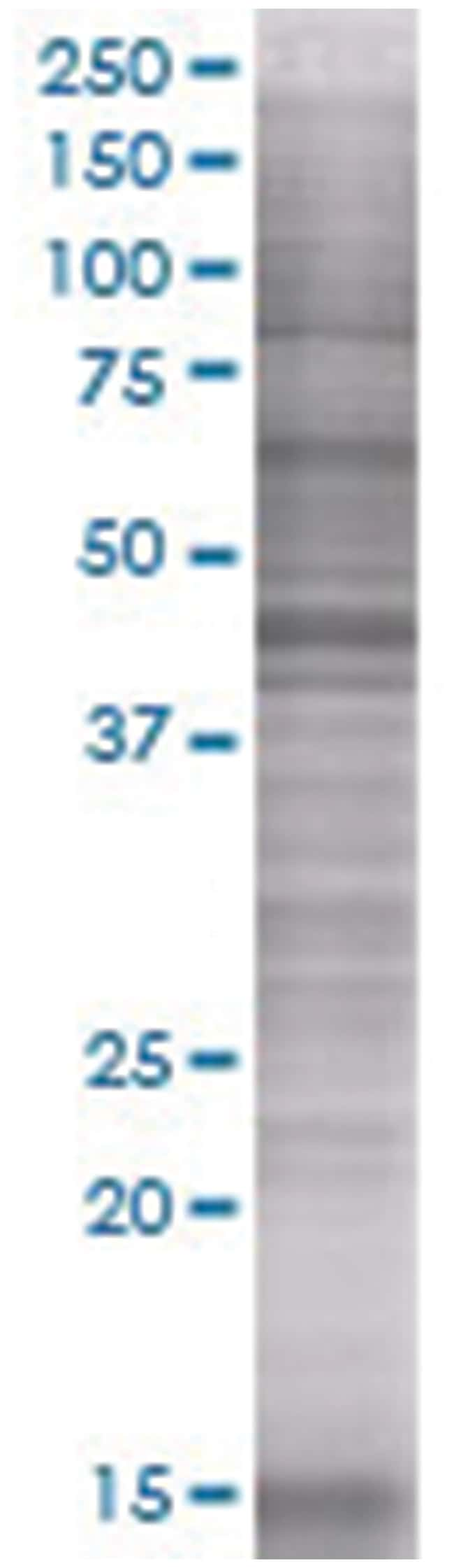
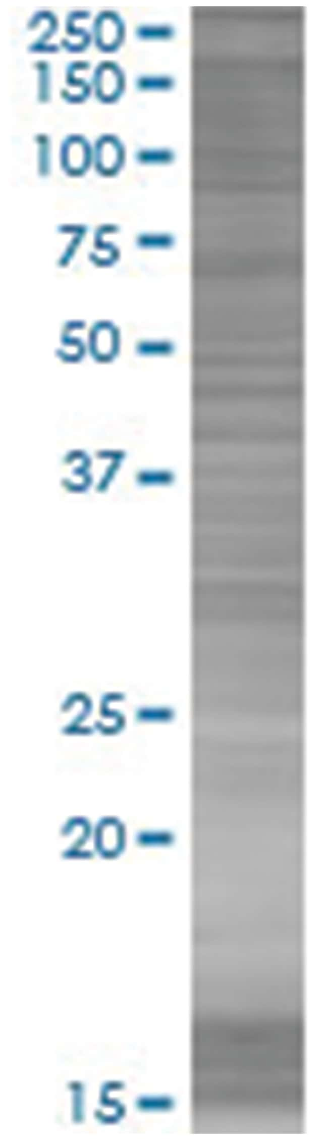
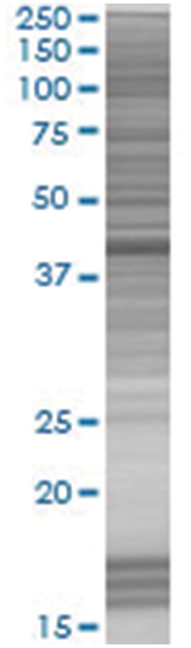
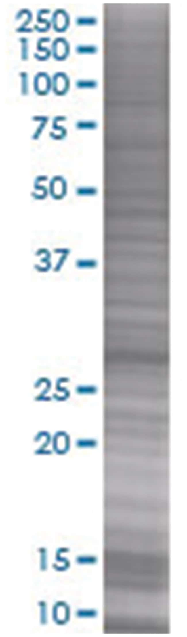
Test du contrôle qualité12.5% SDS-PAGE Stained with Coomassie Blue.
Quantité50 μg
Tampon de stockageIn ready-to-use 1X Sample Buffer (50 mM Tris-HCl, 2% SDS, 10% glycerol, 300 mM 2-mercaptoethanol, 0.01% Bromophenol blue).
Espèces hôtesMouse
Contenu et stockageStore at -80°C. Aliquot to avoid repeated freezing and thawing.
Une question, une configuration particulière ?
Contactez-nous pour un devis.